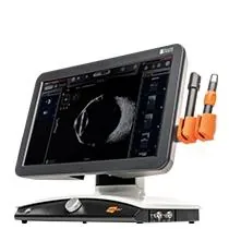


Discover our ultimate ultrasound platform that offers unmatched image quality with an exceptional level of detail.
UBM is a high-frequency ultrasound imaging technique used to explore the anterior segment of the eye, including structures behind the iris. UBM typically uses ultrasound frequencies between 35 and 100 MHz, achieving an axial resolution of approximately 20–50 microns, allowing visualization of microstructures not accessible with standard ultrasound or OCT.
It helps clinicians better understand eye anatomy, diagnose diseases, and guide treatment decisions, especially when standard exams (slit lamp, gonioscopy, OCT) are not sufficient.
Unlike optical imaging technologies, this technique uses high-frequency ultrasound to penetrate pigmented tissues and opaque media. This allows visualization of areas located behind the iris, including the ciliary body, zonules, posterior chamber, and lens equator. Ultrasound Biomicroscopy is particularly valuable when clinicians need to:
The ability to visualise beyond the iris makes UBM an indispensable complement to OCT of the anterior segment.
One of the most common applications of Ultrasound Biomicroscopy is the evaluation of angle-closure disease.
While gonioscopy remains the gold standard for angle assessment, UBM provides additional anatomical information that aids clinicians identify the underlying mechanism of closure.
Studies have shown that UBM can identify the mechanism of angle closure in up to 80–90% of cases, particularly when gonioscopy is inconclusive.*
UBM can assist in the diagnosis and management of:
By visualizing the relationship between the iris, ciliary structures, and lens, UBM enables clinicians to distinguish between different mechanisms that may appear similar during routine examination.
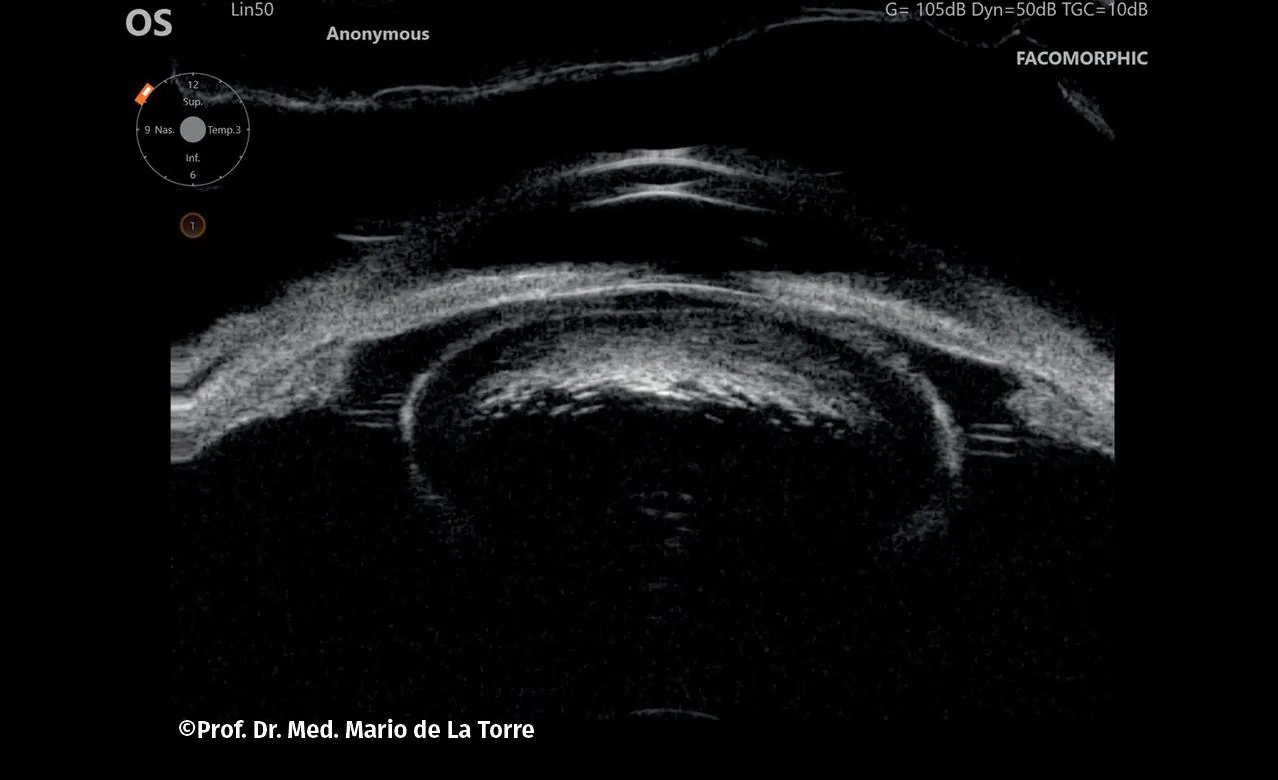
Content to come.
Content to come.
Content to come.
Content to come.
Text d’introduction de cette section. Lorem ipsum dolor sit amet, consectetur adipiscing elit, sed do eiusmod tempor incididunt ut labore et dolore magna aliqua. Ut enim ad minim veniam, quis nostrud exercitation ullamco laboris nisi ut aliquip ex ea commodo consequat.
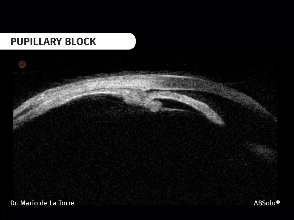
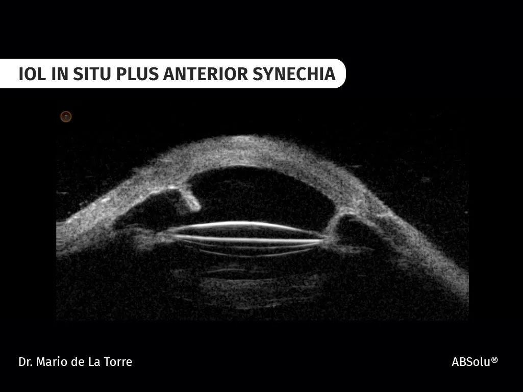
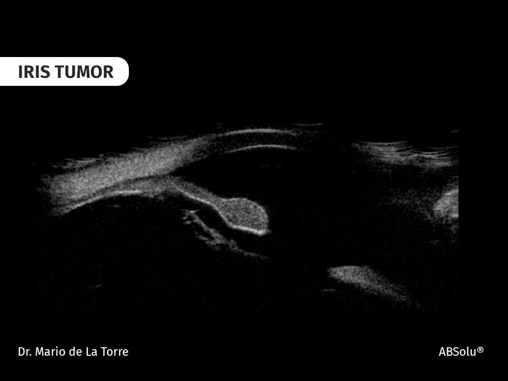
Many ophthalmologists are initially intimidated by Ultrasound Biomicroscopy. The exam is often perceived as operator-dependent and more technically demanding than other anterior segment imaging modalities. However, experienced users agree that acquiring clinically useful UBM scans is easier than many clinicians expect.
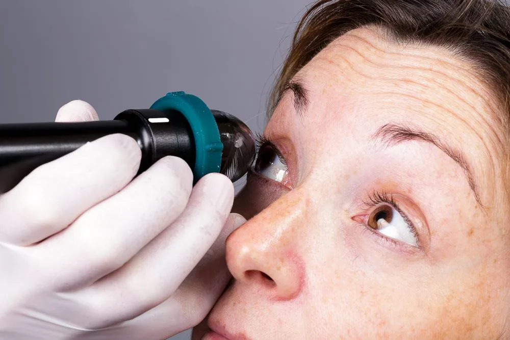
Before the exam, define your goal:
A clear objective improves both image acquisition and interpretation.
Accurate diagnosis starts with recognizing normal structures:
Once these are well identified, abnormalities are easier to detect.
Always scan the four quadrants:
This ensures consistency and reduces the risk of missing findings.
Too much probe pressure can distort anatomy and create misleading images.
With experience, clinicians quickly learn how little pressure is actually required.
Aim for gentle, stable contact to preserve natural structures.
Do not rely on a single scan:
Small adjustments can greatly improve results.
UBM should always be interpreted alongside:
UBM often explains anatomical findings not visible clinically.
Start with common indications:
Practice and case comparison quickly improve confidence.

Prof Mario de La Torre, Lima (Peru)
"Don't try to identify pathology first. Learn to recognize normal anatomy consistently, and pathology will become obvious"
This principle remains one of the most valuable lessons shared by experienced UBM users and educators.
Text d’introduction de cette section. Lorem ipsum dolor sit amet, consectetur adipiscing elit, sed do eiusmod tempor incididunt ut labore et dolore magna aliqua. Ut enim ad minim veniam, quis nostrud exercitation ullamco laboris nisi ut aliquip ex ea commodo consequat.
UBM becomes easy with practice, structure, and repetition.
Text d’introduction de cette section. Lorem ipsum dolor sit amet, consectetur adipiscing elit, sed do eiusmod tempor incididunt ut labore et dolore magna aliqua. Ut enim ad minim veniam, quis nostrud exercitation ullamco laboris nisi ut aliquip ex ea commodo consequat.
Discover our ultimate ultrasound platform that offers unmatched image quality with an exceptional level of detail.
While anterior segment OCT provides axial resolutions of approximately 5–10 microns, its penetration is limited by pigment and opaque media. In contrast, UBM (35–50 MHz) allows deeper penetration through pigmented tissues, although at lower resolution.
Key differences:
| Feature | UBM | OCT |
|---|---|---|
| Technology | High-frequency ultrasound | Light-based imaging |
| Penetration | Excellent (through iris & opacity) | Limited by pigmentation/media opacity |
| Resolution | 20–50 µm | 5–10 µm |
| Best for | Ciliary body, hidden structures | Cornea, angle, superficial structures |
| Contact | Yes (immersion probe) | No |
Clinical takeaway:
Answer to come.
Answer to come.
Answer to come.
Answer to come.
Answer to come.
Answer to come.
Answer to come.
Answer to come.
References :